

The size and prominence of llama eyes is one of the first things that people notice and is part of what makes them so endearing. Llama eyes are only slightly smaller than cattle or horse eyes, but they are placed in a much smaller head. The prominence of their eyes does make trauma and damage to the cornea one of the more common ocular problems in llamas. Signs of corneal disease include excessive tearing and blinking, holding the eye partially closed, white/gray areas on the eye, conjunctivitis, pain or discomfort and/or purulent discharge (pus) coming out of the eye. If you see any of these signs contact your veterinarian for treatment.
 |
Tear production in llamas is slightly different than most other species. The tear film normally has three layers: a deep mucous layer right against the eye, a middle aqueous (fluid) layer, and an outer oil layer. The mucous layer is produced by cells (goblet cells) which are along the conjunctiva. This layer holds the aqueous layer against the eye and has some antibacterial activity. The aqueous layer is produced by the lacrimal gland and the gland of the third eyelid. It provides most of the moisture and performs most of the flushing action of the tears. The outer oil layer comes from the tarsal (meibomian) glands which line the edges of the eyelids in most species. Camelids do not have tarsal glands and their oil layer comes from a sebaceous gland (another type of oil secreting gland) on the lacrimal caruncle (the triangular lump of tissue in the inside corner of the eye). The main function of the oil layer is to increase surface tension and prevent evaporation of the aqueous layer.
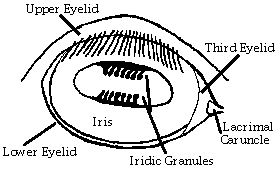
The iris of camelids is also unique. Like other species, the iris can be a range of colors from dark brown to blue (non-pigmented). There may be specks of pigment in blue eyes and specks of blue in pigmented eyes. Like all other ruminants, the pupil is oval shaped with the horizontal axis being the longer one. The difference comes with structures called iridic granules or corpora nigra. These are dark lumpy masses coming off the iris at the top and/or bottom of the pupil margins. They are thought to shade the eye from bright sunlight. Horses only have these on the top pupillary margin and they are more rounded. Cattle and sheep have more rounded iridic granules like horses, but on both sides (top and bottom) of the pupil. Llamas have iridic granules on the top and bottom of the pupil as well, but they are more elongated than rounded. In bright sunlight these iridic granules can actually interdigitate to completely cover the center of the pupil. This leaves just two holes open on either end of the pupil. Take a look at your llama's eyes. These structures are prominent and can easily be seen with the naked eye.
 |
The fundus of the eye is basically the back of the eye where the retina lies. This area of the eye can only be seen by dilating the pupil and using an ophthalmoscope or magnifying glass. The retina lies over a dark non-reflective portion and a colored reflective portion (in most species) along with the optic disk (area where the optic nerve comes into the eye). The colored reflective portion of the fundus is called the tapetum lucidum. It is used to reflect light back into light receptors of the retina and improve night vision. The reflection of the tapetum is what you see when headlights shine into animals' eyes at night. Llamas are not usually active at night and thus, do not have a tapetum.
The fundus also has a characteristic pattern of blood vessels for different species. The camelid pattern of vessels is very similar the pattern for cattle. There are three main pairs of vessels which leave from the optic disk area and radiate out.
Last updated February 17,1997
 |
 |